Digestive System Labeled: What Does Each Organ Do?
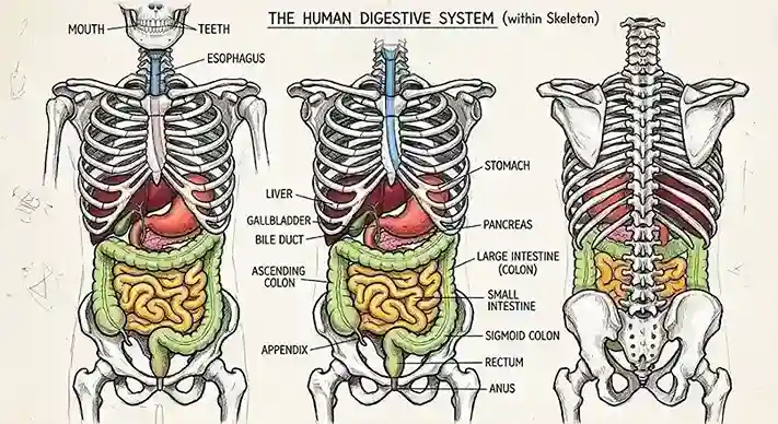
Staring at a textbook diagram and still unsure where the stomach ends and the small intestine begins? You are not alone. Most students and parents struggle to match organ names to their shapes, and the confusion grows when the labels crowd together. This guide gives you a clear labeled digestive system map, explains what every part does, and shows you how to remember them.
What Is the Digestive System?
The digestive system breaks down the food you eat into small molecules your body can absorb. It also removes waste. Two groups of organs make this happen: the alimentary canal, which is a continuous tube, and the accessory organs, which release enzymes and fluids through ducts.
Digestion starts before you swallow. Saliva begins breaking down starch as you chew. From there, a series of organs takes over, and each one adds a specific chemical or mechanical action.
Labeled Digestive System Diagram: Every Organ at a Glance
Use this table as your reference when you study a labeled digestive system chart. Each row names the organ, places it in the body, and gives its main role.
| Organ | Location | Main Job |
| Mouth | Head, front of face | Chews food and mixes it with saliva |
| Pharynx | Behind mouth and nose | Passes food from mouth to esophagus |
| Esophagus | Runs through the chest | Moves food down to the stomach |
| Stomach | Upper left abdomen | Stores food and mixes it with acid |
| Liver | Upper right abdomen | Produces bile and processes nutrients |
| Gallbladder | Under the liver | Stores and releases bile |
| Pancreas | Behind the stomach | Releases digestive enzymes and insulin |
| Small intestine | Central abdomen | Digests food and absorbs most nutrients |
| Large intestine | Frames the small intestine | Absorbs water and forms stool |
| Rectum | Lower pelvis | Stores waste before elimination |
| Anus | End of the tract | Controls the release of waste |
The Alimentary Canal: Mouth to Anus
The alimentary canal is one long tube, roughly 9 meters (about 30 feet) in an adult body. Food moves through it in a fixed order. Learn this sequence and the labeled digestive system diagram becomes much easier to read.
- Mouth: Teeth crush food, and salivary enzymes start breaking down starch.
- Pharynx: This shared passage carries food past the throat and into the esophagus.
- Esophagus: A muscular tube that pushes food toward the stomach in about seconds.
- Stomach: A J-shaped sac that churns food and exposes it to strong acid and the enzyme pepsin.
- Small intestine: Divided into the duodenum, jejunum, and ileum, where most digestion and absorption occur.
- Large intestine: Includes the cecum, colon, and rectum, where water is reclaimed and waste is compacted.
- Anus: The final exit point, controlled by ring-shaped muscles called sphincters.
Accessory Organs That Support Digestion
Accessory organs do not sit inside the food tube, yet they are essential. They pour chemicals into the canal through small ducts. Without them, digestion would stall.
The liver produces bile, a fluid that breaks up fats into tiny droplets so enzymes can work on them. The gallbladder stores bile between meals and releases it when fatty food arrives. The pancreas secretes powerful enzymes for proteins, fats, and carbohydrates into the duodenum. The salivary glands in the mouth start the process before food reaches the stomach.
How Food Moves Through the Tract
Your body does not rely on gravity to move food. Smooth muscles contract in waves, a process called peristalsis. These waves push food forward even when you lie down or stand on your head.
Follow the journey in this order:
- You chew and swallow, forming a soft ball called a bolus.
- Peristalsis moves the bolus down the esophagus into the stomach.
- The stomach churns the food into a thick liquid called chyme.
- Chyme enters the small intestine a little at a time.
- Bile and pancreatic enzymes finish breaking down nutrients.
- The intestinal lining absorbs nutrients into the blood and lymph.
- Leftover material moves into the large intestine for water recovery.
Digestion Versus Absorption: Know the Difference
Students often mix up these two terms, and exam questions love to test the difference. Digestion means breaking large food molecules into smaller ones. Absorption means moving those small molecules across the intestinal wall into the bloodstream.
Digestion happens along the whole tract, but absorption concentrates in specific places. The stomach absorbs very little, most of it happening in the small intestine and some in the colon. Keep this distinction in mind when you read any labeled digestive system chart.
The Role of Enzymes and Bile
Enzymes are proteins that speed up chemical reactions. Each one acts on a specific nutrient. Amylase breaks down starch, pepsin breaks down protein in the stomach, and lipase breaks down fats in the small intestine.
Bile is different. It is not an enzyme. It acts like a detergent, emulsifying fats so that lipase can reach them. Without bile, a large share of dietary fat would pass through you unabsorbed.
Where Most Nutrients Are Absorbed
The small intestine does most of the absorbing, and its design explains why. The inner lining forms folds, and these folds bear millions of tiny finger-like projections called villi. Each villus is covered with microvilli, which together create a surface area roughly the size of a tennis court.
Villi contain blood vessels and lymph vessels. Sugars and amino acids enter the blood, while most fats enter the lymphatic system. This large, folded surface is one of the most efficient absorption systems in the body.
The Large Intestine and the Gut Microbiome
The large intestine is shorter than the small intestine, but its width gives it its name. Its main job is to absorb water and salts. It also houses trillions of bacteria that ferment fiber the body cannot digest on its own.
These microbes produce short-chain fatty acids and some vitamins. Eating a varied diet with plenty of fiber supports a balanced community of gut bacteria. This link between diet, microbes, and digestion is a key reason the large intestine matters beyond waste removal.
Common Digestive Problems to Recognize
Knowing the anatomy helps you understand what goes wrong when problems arise. Problems often show up in the organ that performs the failing task.
- Acid reflux: Stomach acid flows back into the esophagus, causing a burning sensation.
- Gallstones: Hardened bile material forms in the gallbladder and may block its duct.
- Constipation: Stool stays in the colon too long, which makes it hard and dry.
- Irritable bowel syndrome: A functional disorder that changes bowel habits and causes cramping.
- Lactose intolerance: The small intestine makes too little lactase to break down milk sugar.
If symptoms persist, a doctor can assess the cause. Self-diagnosis based on a diagram is not reliable.
How to Study a Labeled Digestive System Diagram
Memorizing a labeled digestive system chart works best when you connect each part to a sequence. Try these methods:
- Trace the path with your finger: Start at the mouth and say each organ aloud as you move down the page.
- Draw it from memory: Sketch the tract and the accessory organs, then check your drawing against the original.
- Use a memory phrase: Build a sentence where each word starts with the same letter as each organ.
- Quiz yourself on function: Cover the labels and ask what each organ does, not only where it sits.
Repeat this practice over several days. Spaced review locks the sequence into long-term memory far better than cramming the night before a test.
Tips for Keeping Your Digestive System Healthy
A diagram shows structure, but daily habits determine how well each organ performs. Small changes help the entire system work smoothly.
- Eat fiber from vegetables, fruits, beans, and whole grains.
- Drink water throughout the day to keep stool soft and moving.
- Chew slowly, which gives saliva more time to start digestion.
- Move your body regularly, since physical activity supports bowel motility.
- Limit highly processed foods and large, fatty meals that strain the liver and gallbladder.
- Schedule a checkup if you notice blood in stool, sudden weight loss, or persistent pain.
Put This Knowledge to Work
Now you can read any labeled digestive system diagram with confidence. Start by tracing the path from mouth to anus, then name the accessory organs and their jobs. Print the table in this guide, quiz yourself tonight, and share it with a classmate or family member who needs it too. Leave a comment below with the organ you found hardest to remember, and I will explain its role in a short follow-up guide.